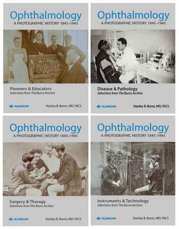
Ophthalmology: A Photographic History 1845-1945
Ophthalmology: A Photographic History 1845-1945, the volumes in this series are individual books on specific topics; however, they are interrelated, and many topics of the volumes are referenced in the other volumes. For example, retinal disease is documented in volume one, Pioneers & Educators, by photomicrographic views and Dimmer’s fundus photographs; in volume two, Disease & Pathology, Bedell’s fundus images; in volume three, Surgery & Therapy, Deutschmann’s retinal procedure; and in volume four, Instruments & Technology, by Dimmer’s fundus camera development. The four volumes contain a total of 218 photographs, plus 18 images on the end pages.
Volume one, Pioneers & Educators, documents some of the leaders of ophthalmic and medical science who laid the foundations of the profession. Numerous portraits are presented; among the notables are Hermann von Helmholtz, Louis émile Javal, William Thomson, Charles Shannon, William Norris, Algernon Reese, Albrecht von Graefe, Cornelius Donders and karl koller. The creation of ophthalmic hospitals is illustrated by images of New York institutions. Some photographs illustrate the advance of medical education and teaching showing ophthalmologists at work. Several photomicrographs and fundus images document the development of retinal histology and photography. The important role of Vienna in ophthalmic education is elucidated through images and documents. Photographs by A. de Montméja, published in his 1870s journal, illustrate ophthalmic conditions of the era.
Volume two, Disease & Pathology, presents unusual case studies and ophthalmic conditions. The book starts with the earliest surviving images, daguerreotypes, of patients with ocular disease. Dramatic photographs of patients su ering from a wide range of ophthalmic conditions include: retinal detachment, neoplasms, genetic disease, and infections from syphilis, tuberculosis, trachoma, and ophthalmia neonatorum. Trauma images include the rst presentation of ocular gunshot wound with several previously unpublished views of wounded soldiers in the Civil War. Images from Victor Morax’s private photograph album document the ‘father of ophthalmic bacteriology.’
Volume three, Surgery & Therapy, contains many iconic images of the development of ophthalmology. The end papers of each volume show the hand-colored images of Paris’ Julius Sichel. The images are pre- and post- operative examples of an arti cial pupil. Photographs of pioneer surgeons, patients and procedures document the development of cataract, retinal and glaucoma surgery. A series of photographs of cataract and other procedures portray the change in dress of the operators, patients and level of sterility and anesthesia. Techniques in x-ray diagnosis and radiation therapy are also illustrated. Numerous therapies of ocular disease, from the most ancient (bloodletting) to images of several physical therapies, enable us to recognize the frustration in treating ocular disease and realize the phenomenal advances in treatment.
Volume four, Instruments & Technology, documents experiments, personalities and the development of instrumentation that de ned the profession. It clearly illustrates the increasing sophistication and development of ophthalmology’s tools. Ophthalmologists’ ability to diagnose disease superseded ability to treat disease. This book describes the development and/or use of several key ophthalmic instruments, including the ophthalmoscope, sideroscope, keratometer, refractive devices, stereoscope, fundus camera, tonometer, eikonometer and phoropter.
256 pages | 4 Hardcover Volumes in Slipcase | 11.5 x 8.5 inches | ISBN: 978-1-936002-02-3 | 2009


